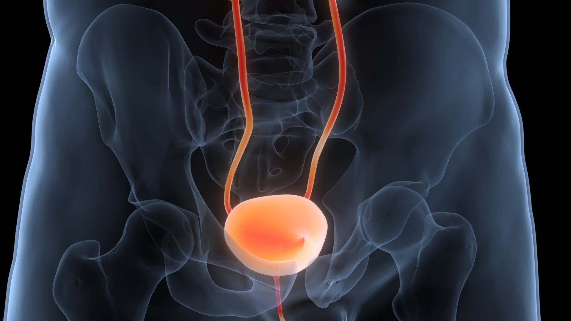
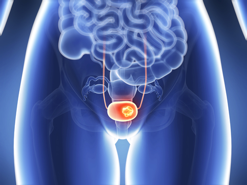
A study published in Clinical Radiology aimed to develop a radiomics model to predict the muscle-invasive status of bladder cancer (BC) in contrast-enhanced computed tomography (CECT) images, compared with radiologists’ interpretations. The analysis found that the model “outperformed the radiologists’ visual assessment in predicting the muscle-invasive status of BC in the venous phase of CT images.”
In this study, 188 CECT images with histopathologically confirmed BC were retrieved, and retrospectively analyzed from November 2018 to December 2019. Radiomics analysis included radiomics feature extraction and model development. Diagnostic performance of radiomics was assessed using receiver operating characteristic (ROC) curve analysis and the area under the ROC curve (AUC).
According to the results, the radiomics model reached an AUC (95% confidence interval [CI]) of 0.979 (0.935-0.996) and 0.894 (0.796-0.956) in both the training and test dataset, respectively. The researchers observed that the radiomics model outperformed the visual assessment of radiologist A and B both in the training (0.865 [0.791-0.921], 0.894 [0.824-0.943]), and test dataset (0.766 [0.647-0.860], 0.826 [0.715-0.907]).
Keywords: bladder cancer, radiologist