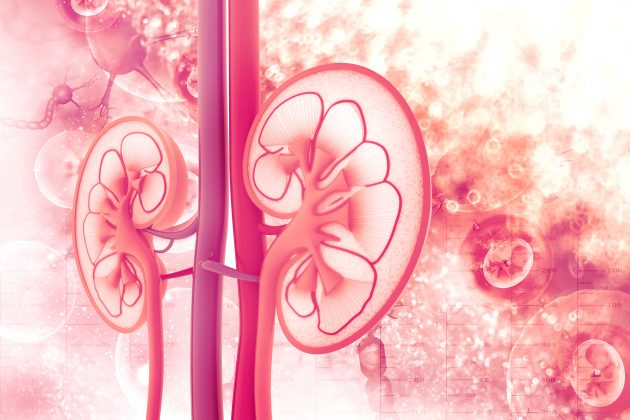
M. Herrmann and colleagues at University Hospital Erlangen, Erlangen, Germany, conducted a study to assess a new diagnostic approach to human leukocyte antigen (HLA) typing in kidney transplant recipients. The approach utilizes cultured renal tubular cells from urine samples to facilitate clinical assessment of donor specificity of de novo occurring HLA antibodies. Results of the assessment were reported during a virtual poster session at the American Transplant Congress 2020 in a poster titled A New Diagnostic Approach to HLA Testing in Kidney Transplant Patients Using Renal Epithelial Cells.
The researchers collected urine samples from 10 patients following allogenic kidney transplantation and a primary culture of renal tubular cells was established according to a published protocol (Zhou et al. 2012, Nature protocols). Following 2 to 4 weeks of tissue culture and the third passage of cells, DNA extraction was performed from a 10 cm2 culture dish.
To validate the analysis, patients were selected for the existence of stored spleen tissue. Only kidney transplant recipients without residual urine excretion were included in order to avoid contamination with patients’ autologous renal cells. HLA typing compared DNA from tubular cells to DNA from spleen, both derived from the donor. Potential contamination of urine samples with patients’ autologous renal cells was further ruled out by performing fluorescence in situ hybridization (FISH) of tubular cultures, in addition to analysis for genetic polymorphic markers by human multiplex short tandem repeats.
Most attempts to establish a culture of renal tubular cells were successful; only rarely did microbial contamination or insufficient proliferation of cells require a repeat urine sample. Use of FISH or analysis of polymorphic markers successfully ruled out relevant contamination with autologous tubular cells. In all patient samples, there was a consistent match in comparison of HLA typing from cultured renal tubular cells with their respective control samples from the spleen. The researchers were able to clinically interpret the findings in the context of circulating HLA bodies in selected cases.
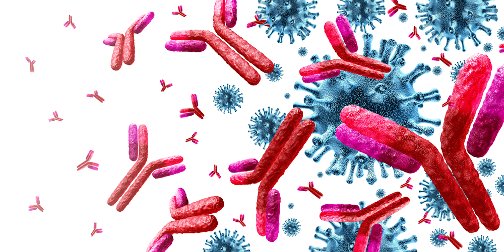
In summary, the researchers said, “Screening for HLA antibodies had broadly become a yearly routine after kidney transplantation. Detection of previously unidentifiable de novo HLA antibodies is common during the follow-up after kidney transplantation due to widespread use of highly sensitive single antigen solid phase tests specific for individual HLA alleles. Assessing donor specificity of these antibodies, however, is frequently limited by incomplete or unavailable donor HLA typing data and/or donor samples for retyping.
“Theoretically, a graft biopsy could deliver donor DNA for HLA typing. However, this avenue is not frequently used since it is an invasive procedure and the acquired tissue is heterogeneous with varying degrees of recipient cellular material. We suggest that this new diagnostic approach for HLA typing in kidney transplantation patients offers a non-invasive option to determine donor specificity of de novo HLA antibodies typically occurring many years after transplantation. We propose that this technique is valuable in clinical management of humoral allograft rejection.”
Source: Herrmann M, Bach C, Knaup KX, Schiffer M, Spriewald B, Wiesener MS. A new diagnostic approach to HLA testing in kidney transplant patients using renal epithelial cells. Abstract of a poster presented at the virtual American Transplant Congress 2020 (Abstract A-283), May 30, 2020.
Credit: Original article published here.