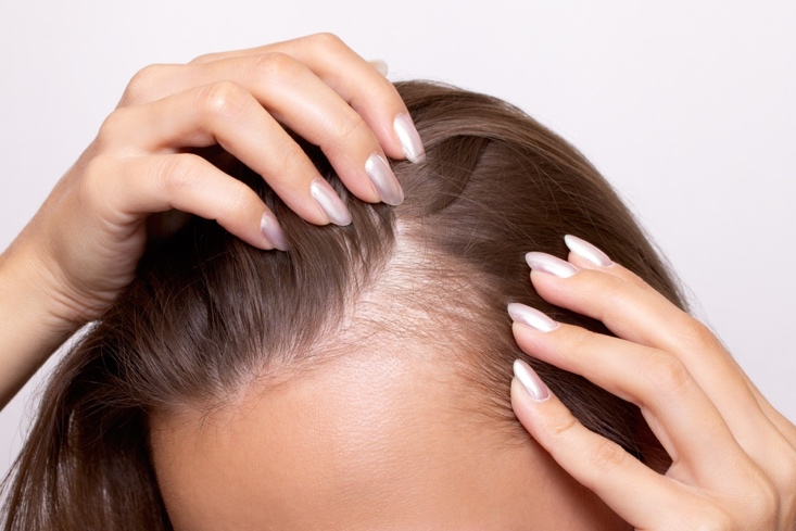
A recent narrative review summarized what’s known about eye involvement in alopecia areata (AA)—from shared immune mechanisms to the spectrum of ocular findings reported from 1963 through February 2025—and offers practical screening guidance for dermatology practice. The proposed link centers on “immune privilege”: both the anagen hair follicle and key ocular tissues actively suppress inflammation. AA-related immune-privilege collapse (plus genetic susceptibility, systemic inflammation, atopy/autoimmune comorbidity, and possible non-immune triggers like oxidative stress or neurogenic inflammation) may help explain ocular comorbidities. Treatment can also contribute (e.g., corticosteroid-associated cataract/glaucoma risk; prostaglandin analog–related ocular surface effects).
Across heterogeneous studies, commonly reported issues include eyebrow/eyelash loss (madarosis), blepharitis, allergic-type conjunctivitis, dry eye/keratitis, and lens changes (often asymptomatic opacities and cataracts), with more variable posterior findings (choroid/RPE/retina). Because the evidence is mixed and confounding is common, the authors recommend routine symptom screening at AA visits using broad questions plus targeted red-flag prompts, with optional PROs to capture burden. For mild ocular surface symptoms, they suggest conservative first-line care (lid hygiene, preservative-free tears, environmental measures). They also recommend early ophthalmology referral for red flags, posterior symptoms, or when starting systemic AA therapy to establish a baseline and monitor for complications.
Reference: Guavita Falla PM, Buendía-Castaño D, Hermosa-Gelbard Á, et al. Ophthalmologic Comorbidities in Alopecia Areata. J Clin Med. 2025 Nov 27;14(23):8409. doi: 10.3390/jcm14238409. PMID: 41375713; PMCID: PMC12693123.