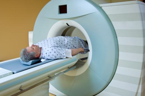
A new study shows that magnetic resonance imaging (MRI) often underestimates the size of prostate tumors, which may impact treatment. The study was published in The Journal of Urology.
In this study, researchers analyzed 461 lesions in 441 men with biopsy proven prostate cancer. They noted that the difference between radiologic tumor size and pathological tumor size was assessed, and clinical, pathological and radiographic predictors of pathological tumor size were examined.
According to the study results, MRI frequently underestimates the size of tumors in prostate cancer, and such underestimation occurs most often when the MRI-measured tumor size is small and the PI-RADS score, which is used to classify lesions in prostate MRI analysis, is low.
The investigators noted that for prostate tumor treatments to be successful, both the MRI size measurement and PI-RADS score must be accurate because they allow physicians to determine precisely where tumors end and where the normal, healthy tissue surrounding them begins.
“Multiparametric magnetic resonance imaging frequently underestimates pathological tumor size and the degree of underestimation increases with smaller radiologic tumor size and lower PI-RADSv2 scores,” the researchers wrote in conclusion.
“Therefore, a larger ablation margin may be required for smaller tumors and lesions with lower PI-RADSv2 scores. These variables must be considered when estimating treatment margins in focal therapy.”
Credit: Original article published here.