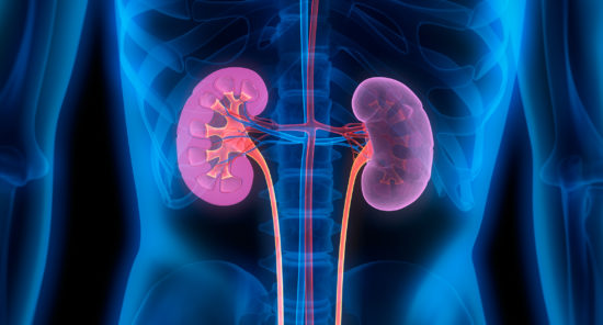
Kidney Week 2020
There is a well-established association between type 2 diabetes and increased kidney volumes; diabetes is also associated with the development of diabetic nephropathy. The UK Biobank cross-sectional study aimed to examine 100,000 subjects ages 44 to 82 years using magnetic resonance imaging (MRI). The resulting images allow measurement of kidney parenchymal volume (KPV).
Lars Johansson and colleagues conducted a study to quantify KPV and examine the association with age in patients with and without type 2 diabetes. Results of the study were reported during a virtual poster session as ASN Kidney Week 2020 in a poster titled The Aging Kidney: Renal Parenchymal Volumes from MRI, a Comparison Between Type 2 Diabetes and Non-Type 2 Diabetes in 37,450 UK Biobank Participants.
The researchers developed and validated an automated deep learning-based method for direct KPV segmentations and measurements that was applied to the UK Biobank MRIs of 37,540 subjects. KPV was analyzed as a function of diagnosed type 2 diabetes (defined as diagnosis of diabetes after the age of 40 years), sex, and age. Correction for lean tissue volumes assessed by MRI was performed in all subjects.
Of the 37,450 individuals, 47.6% were male. In subjects without type 2 diabetes, there was a steady decline in KPV in both males and females. In those with type 2 diabetes (1126 males, 5530 females), KPV is significantly larger, over 50 years of age, compared with those without type 2 diabetes. There is a faster KPV decline in those with type 2 diabetes than in those without type 2 diabetes, Following adjustment for lean tissue volumes from MRI, there was no change in the difference in decline rates between those with and those without diabetes.
In conclusion, the researchers said, “Type 2 diabetes subjects have a larger KPV than non-type 2 diabetes in middle-aged subjects but show a faster KPV decline, independent of lean tissue volume differences. The faster decline in type 2 diabetes can potentially be explained by increased hyperfiltration and oxidative stress in type 2 diabetes.”
Source: Johansson L, Langner T, Hockings P, et al. The aging kidney: Renal parenchymal volumes form MRI, a comparison between type 2 diabetes and non-type 2 diabetes in 37.450 UK Biobank participants. Abstract of a poster presented at the American Society of Nephrology virtual Kidney Week 2020 (Abstract PO0960), October 22, 2020.
Credit: Original article published here.