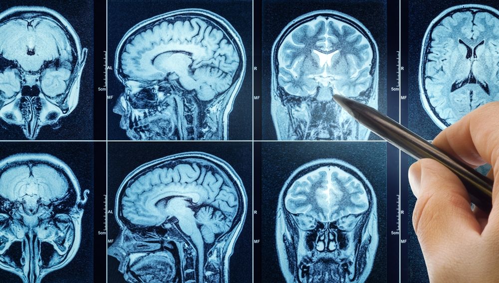
Psychosis—especially visual hallucinations (VH)—impacts up to half of people with Parkinson’s disease (PD) and worsens quality of life and prognosis, yet mechanisms remain unclear. Current accounts point to disrupted attentional control and imbalanced top–down/bottom–up processing across large-scale networks. Researchers tested these ideas using resting-state functional MRI from Parkinson’s Progression Marker Initiative (PPMI) (PD with visual hallucinations [PDVH], PD without visual hallucinations, healthy controls) with replication in Incidence of Cognitive Impairment in Cohorts with Longitudinal Evaluation-PD (ICICLE-PD). They compared global and network-level functional connectivity (FC) and applied network-based statistics (NBS) to detect hallucination-linked subnetworks, controlling for motion, demographics, clinical covariates, and multiple comparisons.
Global and most canonical network FC differences were nonsignificant after correction, but NBS revealed a reproducible subnetwork of reduced FC in PDVH spanning dorsal/ventral attention, default-mode, and somatomotor regions. In ICICLE-PD, mean FC within this PPMI-derived subnetwork was lower in PDVH and correlated with hallucination severity, though de novo NBS in the full replication sample was negative. In PPMI, lower subnetwork FC related to worse attention and global cognition longitudinally and to higher future motor severity. Findings support an attentional–default mode network (DMN) dysconnectivity signature—potentially shaped by thalamic/basal-ganglia pathways—rather than generalized DMN hyperconnectivity. Despite modest samples, cortical-only analyses, and limited hallucination phenotyping, results support network-informed biomarkers and interventions for PDP.
Reference: Montagnese M, Mehta MA, Ffytche D, et al. Disrupted functional brain network associated with presence of hallucinations in Parkinson’s disease. Brain Commun. 2025;7(3):fcaf185. doi: 10.1093/braincomms/fcaf185.