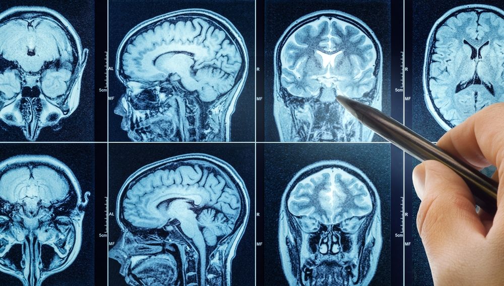
Researchers of this study explored whether gray matter volume loss in Parkinson’s disease psychosis (PDP) is associated with brain expression of genes coding for cannabinoid receptors CB1 and CB2. Using data from a prior coordinate-based meta-analysis of 10 structural MRI studies, the authors compared gray matter volume differences between 211 patients with PDP and 298 patients with Parkinson’s disease without psychosis. Prior results showed that patients with PDP had lower gray matter volume in parietal, occipital, and temporal brain regions after adjustment for Parkinson’s disease medication use and cognitive status.
The analysis also found a significant association between regional gray matter volume loss in PDP and expression of the CB1 receptor gene, even after accounting for levodopa equivalent daily dose and spatial autocorrelation. No significant association was found for CB2 after correction. The authors suggest that CB1 receptor-related pathways may be involved in the neurobiology of psychosis in Parkinson’s disease, possibly through links to neurodegeneration or altered neurotransmitter regulation, though this remains unproven. They note that future studies using in vivo PET imaging are needed to clarify whether CB1 receptor availability differs in patients with PDP compared with those without psychosis.
Reference: Pisani S, Váša F, Velayudhan L, Bhattacharyya S. Gray Matter Volume Loss in Parkinson’s Disease Psychosis and Cannabinoid Receptor Gene Expression in the Brain. Mov Disord. 2026 Feb 20. doi: 10.1002/mds.70222. Epub ahead of print.
Link: https://movementdisorders.onlinelibrary.wiley.com/doi/10.1002/mds.70222